Turtles
(Order Testudines)
Introduction. Turtles are reptiles with a bony carapace. They are of the order Testudines, also known as Chelonia. DNA phylogentic studies place turtles close to Archosaurs (birds, crocodiles and dinosaurs) (Crawford, Parham, & al, 2014). The clade which includes Testudines and Archosaurs is called Archelosauria. Turtles are further subdivided into Cryptodira which can retract the head directly into the shell, and Pleurodira which fold their necks to the side to protect their heads.
Turtles have diversified little since ancestors of the 348 extant species first appeared 220mya, except for a period when continental coastal margins appeared 30mya increasing the size of their potential habitat (Thomson & Spinks, 2021). No taxonomic distinction is made between turtles, tortoises and terrapins. Turtles have round pupils. As with mudskippers, turtles which are primarily aquatic have had to deal with changes in the focussing properties of the cornea depending on whether they are looking through water or air. This problem has been dealt with in sea turtles by evolving flat corneas which do not refract light.
Present study. 33 species of turtle were studied. All turtles blinked with their lower lids. This was accompanied by globe retraction. In 1 of 7 species of the sub-order Pleurodira and in16 of 26 species of the sub-order Cryptodira, a nictitating membrane, mostly grey/white and opaque, came out of the medial canthus as the globe retracted and the lower eyelid rose, and travelled across the eye. Often the lower lid would rise rapidly and then descend more slowly. Blinks varied greatly in length. The nictitating membrane was often visible pre-blink and then covered the eye during the blink. With drowsiness, the lower lid slowly rose. Most had unilateral blinks (winks). Asynchronous bilateral blinks were seen. Most had 2-3 ridges in the lower lid which pulled apart as the lid rose to cover the eye and the globe retracted; the one closest to the edge of the lid was sometimes so thin as be mistaken for a membrane like the frog’s.

Examples
Freshwater turtle
Suborder Cryptodira
Family Trionychidae
Spiny softshell turtle (Apalone spinifera)
Blink type: Globe retraction with elevation of lower lid
A flurry of winks in the left eye, a few in the right. The upper lids sink into the orbits as the globes retract and the lower lids rise.

Right eye: a) Pre-blink b) Nictitating membrane (arrow) moves out from inner canthus c) Lower lid almost covers the eye. The arrow marks where the upper and Lowe lids meet. The upper lid has dropped a little probably because of globe retraction.
Freshwater turtle
Suborder Pleurodira
Family Chelidae
Saw shelled turtle (Myuchelys latisternum)
Blink type: Globe retraction with elevation of lower lid
Wink then blink played at 10% speed

a) Pre-blink. b) Both eyes are retracted and the right covered. The left eye is still visible. The inferior part of the right lower lid (white arrow) is thicker than the superior part which covers the eye. c) Both eyes are covered by the lower lids. d) and e) The left eye opens. f) Post-blink.

Wink in the same specimen at higher magnification: a) Pre-blink. b), c) and d) Left eye is retracted and both upper (orange arrow) and lower lids (white arrow) are prominent as they sink into the orbit. The thinner superior part of the lower lid has covered the left eye. e) The lower lid is descending. f) Post-blink.
Freshwater turtle
Suborder Pleurodira
Family Chelidae
Red-bellied short-necked turtle (Emydura subglobosa)
Blink: Globe retraction with elevation of lower lid

Swimming underwater. a) Pre-blink. b) Lower lid elevated during a blink.
Freshwater turtle
Suborder Pleurodira
Family Chelidae
Murray River turtle (Emydura macquarii)
Blink: Globe retraction with elevation of lower lid
Eye movement and blink played at 10% speed

a) Pre-blink. b) Maximal blink. The upper lid has lowered slightly due to globe retraction. The eye is covered by the lower lid which comprises two parts: a semi-translucent upper section (grey arrow) and an opaque lower section (white arrow), the lower lid. c) Post-blink.
Upward eye movement and lower lid blink with globe retraction, underwater, played back at 20% speed

Murray River turtle underwater: a) Pre-blink. b) Upper lid sinks as globe retracts. Eye covered by semi-translucent upper section of the lower lid (grey arrow) and opaque lower section (white arrow), the lower lid. c) Maximal blink. d) and e) Lower lid descends. f) Post-blink

Lower lid blink with globe retraction in the right eye: a) Pre-blink showing no evidence of a nictitating membrane. b) Blink involving elevation of the upper and lower segments of the lower lid to the level of the pupil. c) Maximum excursion of the lower lid, covering the eye. The upper and lower segments of the lower lid are separated by a crease.
Freshwater turtle
Suborder Pleurodira
Family Chelidae
Eastern long-necked turtle (Chelodina longicollis)
Blink type: Globe retraction with elevation of lower lid
Blink played at 10% speed.

a) Pre-blink. b) Globes retract and right palpebral fissure narrows. c) and d) Lower lid, comprising thin upper section and thicker lower section, covers the eye. e) Lower lid descends. f) Post-blink.
Lower lid blink with globe retraction in the right eye:
a) Pre- blink showing mainly the upper segment of the lower lid (orange). No nictitating membrane visible. b) Blink involving elevation of the upper and lower segments of the lower lid. Again, no nictitating membrane visible. c) Maximum excursion of the lower lid - covering the eye. The upper and lower segments of the lower lid are marked by a crease.


Right eye of another individual from side. a) Pre-blink. b) Lower half of the eye covered by the lower lid, more opaque in the lower region than the upper. c) Full blink. d) Lower lid starts to descend. e) Lower lid now looks brown, perhaps due to its upper part folding behind the lower part. f) Post-blink
Freshwater turtle
Suborder Pleurodira
Family Pelomedusidae
Cape terrapin or African helmeted turtle (Pelomedusa subrufus nigra)
Blink type: Globe retraction with elevation of lower lid
Blink played back at 10% speed. Globes retract causing upper lids to sink into orbits. Lower lids cover globes and then descend.
Freshwater turtle
Suborder Cryptodira
Family Emydidae
Yellow bellied slider (Trachemys script)
Blink type: Globe retraction with elevation of lower lid.
Blink played at 10% speed.

a) Pre-blink. b) Eye covered with a paler upper section and a more opaque lower section of the lower lid. c) and d) Upper section (grey arrow) and lower section (white arrow) of lower lid descending. e) Upper section of the lower lid has now disappeared behind the lower section. f) Post-blink.
Freshwater turtle
Suborder Cryptodira
Family Emydidae
River cooter (Pseudemys concinna)
Blink type: Globe retraction with elevation of lower lid
Played at 50% speed

a) Pre-blink. b) Eye begins to retract and lower lid rises. c) Eye covered by lower lid, semi-translucent in the upper part, opaque and thicker in the lower part. d) Lower lid starts to fall. e) Upper section of lower lid tucks behind lower section revealing eye. f) Post-blink
Freshwater turtle
Suborder Pleorodira
Family Podocnemididae
Madagascan big-headed turtle (Erymnochelys madagascariensis)
Blink type: Globe retraction, elevation of lower lid and horizontal movement of nictitating membrane
Played at 10% speed

a) Pre-blink. b) Globe retracts and lower lid elevates - again in two parts, a thin upper and thick lower part. c) The thick part of the lower lid descends but eye remains covered by a semi-translucent upper part. d) As this descends, a nictitating membrane can be seen half-way across the pupil withdrawing towards the inner canthus. e) More of the pupil is exposed as the membrane withdraws. f) Post-blink. The key observation here is that there appears to be a horizontal nictitating membrane in addition to the two parts of the lower lid.

d) and e) at higher magnification
Terrestrial tortoise
Suborder Cryptodira
Family Testudinidae
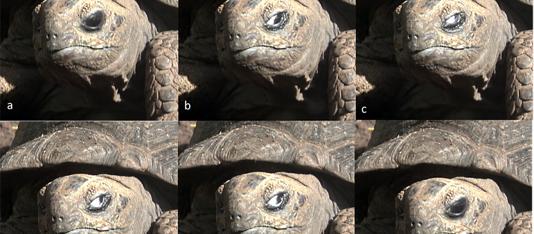
Galápagos tortoise (Chelonoidis niger)
Blink type: Globe retraction, elevation of lower lid and horizontal movement of nictitating membrane
Played at 10% speed

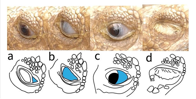
a) Pre-blink. b) Globe retraction begins. c) Full globe retraction causing lids to sink into the orbit. Lower lid elevation, closing the eye. d) Edge of nictitating membrane (grey arrow) visible as eye starts to open. e) Nictitating membrane moves back towards the inner fornix. f) Post-blink, membrane still visible.
Nictitating membrane and lower lid blink with globe retraction, left eye: a) Pre-blink showing a resting nictitating membrane (blue) in the medial canthus. There is a clearly defined lower lid (orange) with no folds. b) Globe retraction and elevation of lower lid with full excursion of nictitating membrane. The upper lid has sunk into the orbit due to globe retraction. c) and d) Lower lid starts to fall and nictitating membrane begins to return to resting position.

Terrestrial tortoise
Suborder Cryptodira
Family Testudinidae
Elongated tortoise (Indotestudo elongata)
Blink type: Globe retraction, elevation of lower lid and horizontal movement of nictitating membrane
Played at 10% speed

Video 1. Blink in an elongated turtle video-ed at 25fps, played back at 30% speed.
At 80ms, the eyeball has retracted and the lower eyelid has elevated, covering the eye. By 400ms, the lower eyelid has descended revealing a nictitating membrane which half covers the eye. Its free edge is at right angles to the palpebral fissure.
Nictitating membrane and lower lid blink with globe retraction, left eye: a) Pre-blink showing the nictitating membrane (blue) in its resting position and the two segments of the lower lid (orange) separated by a crease. b) Partial blink involving the nictitating membrane covering the pupil and elevation of the lower lid. c) Maximum excursion of the lower lid, covering the eye. d) Pre-blink. Sagittal plane of the eye showing the two segments of the lower eyelid, e) The segments of the lower lid start to expand around the cornea. f) The lower lid covers the cornea.

Terrestrial tortoise
Suborder Cryptodira
Family Testudinidae
Aldabra giant tortoise (Geochelone gigantea)
Blink type: Globe retraction, elevation of lower lid and horizontal movement of nictitating membrane
Played at 10% speed

a) Pre-blink. b) Opaque nictitating membrane moves across the eye from the inner canthus. Palpebral fissure narrows probably due to retraction of the globe. Lower lid elevates slightly. c) Maximal excursion of nictitating membrane and lower lid. d) and e) Nictitating membrane withdraws. f) Post-blink.
Terrestrial tortoise
Suborder Cryptodira
Family Testudinidae
Leopard tortoise (Stigmochelys pardalis)
Blink type: Globe retraction, elevation of lower lid and horizontal movement of nictitating membrane
Played at 10% speed

Blink on head turn. a) Pre-blink. b) Retraction of globe, elevation of lower lid, appearance of white nictitating membrane at inner canthus. c) Maximal blink with full retraction of globe. d) Lower lid starting to fall revealing nictitating membrane covering the eye. e) Lower lid down, globe unretracted, nictitating membrane halfway across the eye. f) Post-blink

Nictitating membrane blink on head turn, right eye: a) Pre-blink showing the resting nictitating membrane (blue) in the medial canthus. b) The nictitating membrane has almost covered the eye and with no elevation of the lower lid. c) The head has turned and the nictitating membrane has started to retreat. d) The upper lid has fully risen and the globe has retracted causing the lid to sink into the orbit.
Sea turtle
Family Cheloniidae
Suborder Cryptodira
Loggerhead sea turtle (Caretta caretta)
Blink type: Globe retraction, elevation of lower lid and horizontal movement of nictitating membrane

Still photographs from National Geographic documentary on Youtube: a) Pre-blink. Yellow upper lid. The white area of the eye in the outer canthus is sclera. b) Globe retraction causing upper lid to sink into orbit. c) Glimpse of nictitating membrane (grey arrow) withdrawing into inner canthus as globe unretracts. d) Post-blink.
Sea turtle
Family Cheloniidae
Suborder Cryptodira
Green sea turtle (Chelonia Midas) filmed on Heron Island
Blink type: Globe retraction, elevation of lower lid and horizontal movement of nictitating membrane
Played at 10% speed

a) Nictitating membrane (blue) appears in the medial canthus. b: Nictitating membrane begins to move across the eye as the lower lid rises.
Further comments on sea turtles
The nictitans of the leatherback turtle has multiple folds in the bulbar surface of the membrane probably to increase the production of mucus in order to protect the cornea from the hypertonic salty secretions of the Harderian gland (Brudenall, Schwab, & Fritsches, 2008).
Legler (Legler, 1993) reports that Harderian glands and ducts are absent in all Australian chelids and in all chelonian taxa which lack or have vestigial nictitating membranes. Lachrymal glands occur in all chelonians. They secrete highly concentrated tears in marine turtles and some estuarine turtles. Turtles lack a nasolachrymal duct from the conjunctival cavity to the nasal passageways. Nictitating membranes are absent in the cryptodiran families Kinosternidae, Dermatemyidae and Carettochelydidae, and are absent or rudimentary in many pleurodires of both families. The membrane is absent or vestigial in all Australian chelids.
Mechanism by which the nictitating membrane and lower lid are moved in turtles

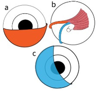
The figure shows the connections of the quadratus muscle in the left eye of a turtle (adapted from Walls 1943).
a) Lower lid (orange) on the front of the eye.
b) Quadratus muscle on the back of the eye with a tendon to the lower lid (orange); and another tendon to the nictitating membrane (blue).
c) Nictitating membrane (blue) on the front of the eye.
Discussion
In most of the freshwater aquatic species studied, blinking involved retraction of the globe and elevation of the lower lid, which usually had an upper and lower part, the upper part being thinner and more transparent. Unlike the nictitating membrane of the frog, which folds under the lower lid, the thin upper segment of the lower lid concertinas back into the lower section.
In all the terrestrial species and the Madagascan big-headed turtle and loggerhead sea turtle, an opaque nictitating membrane moved across the eye from the inner canthus during a blink. There was also retraction of the globe and elevation of the lower lid. Unlike other turtles, marine turtles are said to have mobile upper as well as lower eyelids. In the two studied, the loggerhead and green turtle, the upper lids were certainly prominent but it was hard to say whether they were drawn over the eye actively or sank passively into the orbit as the globe retracted. It looked more like the latter.
Lubrication of the cornea is provided by a lacrimal gland and an Harderian gland with a single duct. There is no nasolacrimal duct (Gans, 1970). In marine turtles, the lacrimal gland is enormously enlarged and excretes a salty solution which can be seen trickling out of the eye when the turtle is on land. The Harderian gland lubricates the eye (Wyneken, 2001).
The lower lid is pulled up from the lateral side of the eye. The nictitating membrane is pulled across from the medial side of the eye by its tendon in the fornix below the eye. As the tendons arise from the same muscle, the lid and membrane have to move together. Simultaneous retraction of the globe increases the degree to which the lid and membrane cover the globe.
It seems that this configuration does not entirely hold for all turtles as the Leopard tortoise (Stigmochelys pardalis) above, was able to cover the eye with the nictitating membrane before elevating the lower lid.
Werneberg found that the pyramidalis muscle was integrated with the retractor bulbi muscle in one specimen of Emydura subglobosa (Werneburg, 2011). Presumably movement of the nictitating membrane would always be accompanied by globe retraction in such individuals.
Innervation
The quadratus and retractor bulbi muscles are supplied by the VIth cranial nerve (Gans, 1970).
References
Brudenall, D., Schwab, I., & Fritsches, K. (2008). Ocular morphology of the Leatherback sea turtle (Dermochelys coriacea). Vet Ophthalmol, Mar-Apr;11(2):99-110. doi: 10.1111/j.1463-5224.2008.00607.x. PMID: 18302574.
Crawford, N., Parham, J., & al, e. (2014). A phylogenomic analysis of turtles. Molecular Phylogenetics and Evolution, 83: 250-257.
Ecker, A (Translated by Haslam, G). 1889. The anatomy of the frog. Oxford: OUP.
Gans, C. (1970). Biology of the reptilia. London and New York: Academic Press.
Legler, J. (1993). Morphology and physiology of the chelonia. In C. Glasby, G. Ross, & P. Beesley, Fauna of Australia.Canberra: AGPS Canberra.
Thomson, T., & Spinks, P. a. (2021). A global phylogeny of turtles reveals a burst of climate-associated diversification on continental margins. PNAS, 118 (7) e2012215118; https://doi.org/10.1073/pnas.2012215118.
Walls, G. (1943). The vertebrate eye and its adaptive radiation. Bloomfield Hills, Mich.: Cranbrook Institute of Science. Cranbrook Press. Bulletin no 19.
Werneburg, I. (2011). The cranial musculature of turtles. Palaeontologia Electronica, Vol. 14, Issue 2; 15A:99p; palaeo-electronica.org/2011_2/254/index.html.
Wyneken, J. (2001). The Anatomy of Sea Turtles.


